Masson's Trichrome Stain Kit
BQC REDOX Technologies
- Catalog No.:
- BQC-KH07007-125
- Shipping:
- Calculated at Checkout
$266.00
Please note that BQC REDOX Technologies items have a $300 minimum. Please contact us if you have any questions.
Purpose of Use:
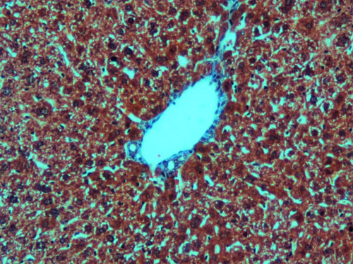
Masson’s trichrome stain is used to demonstrate connective tissue elements, collagen and muscle fibers. After staining, collagen and mucin will be coloured in blue, muscle fibers, cytoplasm and keratin in red and the nuclei blue/black.
Use and sample preparation:
Formalin-fixed, paraffin and frozen samples can be used. After each step, rinsing with tap water and distilled water is required unless specified
Components of Masson’s trichrome stain kit:
- Hematoxylin Weigert’s Reagent A
- Hematoxylin Weigert’s Reagent B
- Phosfotungstic Phosfomolibdic Acid Reagent
- Aniline Blue
- Glacial acetic acid 1%
- Bouin fixative reagent
- Biebrich scarlet
Short procedure:
- Place slide in Bouin’s reagent at 60ºC
- Let the slides cool
- Mix equal parts of Weigert’s Reagent A and B
- Place slide in Weigert’s Hematoxylin reagent freshly prepared
- Apply Briebrich Scarlet and do not rinse
- Apply Phosphotungstic phosphomolybdic acid. Do not rinse
- Apply Aniline blue reagent
- Apply Glacial acetic acid 1%
- Dehydrate with ethanol, clear and coverslip
Stain characteristics:
Collagen and mucin can be visualized as blue, muscle fibers, cytoplasm and keratin in red and the nuclei blue/black.
| Documents & Links for Masson's Trichrome Stain Kit | |
| Manual | Masson's Trichrome Stain Kit Manual |
| Documents & Links for Masson's Trichrome Stain Kit | |
| Manual | Masson's Trichrome Stain Kit Manual |


