")
Exosomes are small vesicles with a diameter of 30 to 200 nm that are secreted from almost all cells that make up the body, and are present in all body fluids such as blood and urine (1)-(5) . They are also secreted in vitro into the culture supernatant of animal cells. Like cells, exosomes are surrounded by a lipid bilayer membrane, with membrane proteins present on their surface and proteins and microRNAs contained inside. It is thought that exosomes are responsible for intercellular communication through the function of these proteins and microRNAs in target cells that have taken up exosomes (6) . One of the structural characteristics of exosomes is the tetraspanin family that is present on the surface. In dogs, CD9 and CD63 are members of this family and exist as surface markers for exosomes (7),(8) .
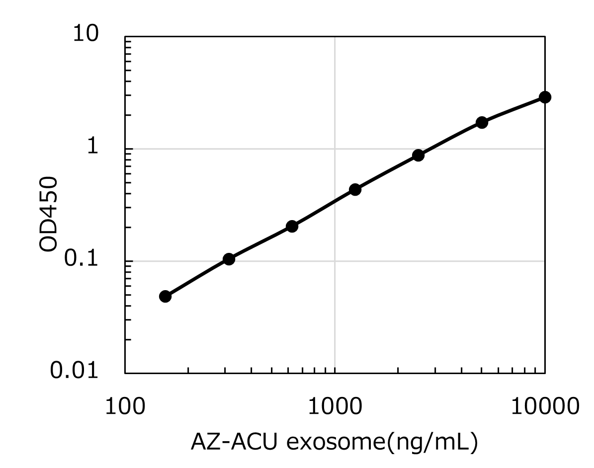
Methods for quantifying exosomes include the amount of protein contained in exosomes and particle analysis using the nanotracking method (9) , but these methods require exosomes to be purified first by ultracentrifugation or other methods. There are very limited means to directly quantify exosomes in body fluids or cell culture media, and no general method has been developed to date.
This kit uses a two-step sandwich ELISA with high-performance antibodies against the exosome markers canine CD9 and CD63, allowing for the relative quantification of exosomes bearing canine CD9 and CD63 molecules on their surface.