Anti ACVRL1 mAb (Clone 8B6)

CUSABIO

SKU:
CSB-RA555022A0HU-50
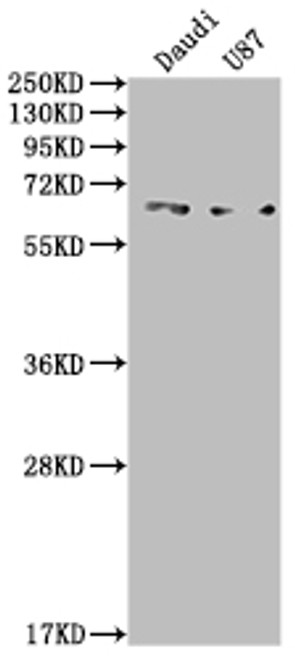
  • Western Blot<br />
Positive WB detected in: Daudi whole cell lysate, U87 whole cell lysate<br />
All lanes: ACVRL1 antibody at 1:2000<br />
Secondary<br />
Goat polyclonal to rabbit IgG at 1/50000 dilution<br />
Predicted band size: 57 kDa<br />
Observed band size: 65 kDa<br />
  • IHC image of CSB-RA555022A0HU diluted at 1:100 and staining in paraffin-embedded human liver cancer performed on a Leica BondTM system. After dewaxing and hydration, antigen retrieval was mediated by high pressure in a citrate buffer (pH 6.0). Section was blocked with 10% normal goat serum 30min at RT. Then primary antibody (1% BSA) was incubated at 4℃ overnight. The primary is detected by a Goat anti-rabbit IgG polymer labeled by HRP and visualized using 0.05% DAB.
Shipping:
Calculated at Checkout
$221.00
Adding to cart… The item has been added
Type I receptor for TGF-beta family ligands BMP9/GDF2 and BMP10 and important regulator of normal blood vessel development. On ligand binding, forms a receptor complex consisting of two type II and two type I transmembrane serine/threonine kinases. Type II receptors phosphorylate and activate type I receptors which autophosphorylate, then bind and activate SMAD transcriptional regulators. May bind activin as well.

Product Specifications
Application WB, IHC, ELISA
Reactivity Human
Clonality Monoclonal (Clone No.: 8B6)


Documents & Links for Anti ACVRL1 mAb (Clone 8B6)
Datasheet Anti ACVRL1 mAb (Clone 8B6) Datasheet
Vendor Page Anti ACVRL1 mAb (Clone 8B6) at CUSABIO

Documents & Links for Anti ACVRL1 mAb (Clone 8B6)
Datasheet Anti ACVRL1 mAb (Clone 8B6) Datasheet
Vendor Page Anti ACVRL1 mAb (Clone 8B6)