Exosomes are small vesicles with diameters of 30 to 200 nm that are secreted by almost all cells that make up the body and are present in all body fluids, including blood and urine (1)-(5) . They are also secreted in vitro into the culture supernatant of animal cells. Like cells, exosomes are enveloped in a lipid bilayer membrane, with membrane proteins present on their surface and proteins and microRNAs encapsulated inside. It is believed that exosomes are responsible for intercellular communication through the function of these proteins and microRNAs in target cells that have taken up exosomes (6) . One of the structural features of exosomes is the tetraspanin family present on their surface. CD9, CD63, and CD81 are members of this family and are also surface markers for exosomes (7) .
Human mesenchymal stem cells (hMSCs) are pluripotent cells with both tissue regeneration and immunoregulatory activities, making them an attractive tool for cell therapy. In recent years, it has been shown that the beneficial effects of hMSCs may be due to paracrine effects and may be mediated, at least in part, by extracellular vesicles (e.g., exosomes) (8), (9) .
Exosome quantification methods include the protein content of exosomes and particle analysis using nanotracking (10) , but these methods require exosomes to be purified by ultracentrifugation or other methods. There are very limited means to directly quantify exosomes in body fluids or cell culture media, and no general method has been developed to date. This
kit is a two-step sandwich ELISA kit that detects CD90-positive and CD63-positive exosomes secreted by human MSCs using high-performance antibodies against CD63, the tetraspanin most highly expressed in hMSCs, and CD90, a proposed MSC-derived exosome marker (11) .
Features
- Direct quantification of CD90+CD63+ exosomes contained in human cell culture supernatants, etc.
- No special equipment is required; measurements can be performed with a regular plate reader (wavelength 450 nm).
- Stability and reproducibility are ensured by using CD90/CD63 fusion protein (standard protein) instead of exosomes themselves, which lack storage stability as a standard reagent.
- Relative quantification of each sample is possible by reading from a standard curve using CD90/CD63 fusion protein (standard protein).
- Detect CD90+CD63+ exosomes using a two-step sandwich method using immobilized CD90 antibody and HRP-labeled CD63 antibody.
What's included
| Contents | capacity | quantity | |
|---|---|---|---|
| 1 | Anti-CD90 antibody immobilized plate | 96well (8well x 12 strips) | 1 sheet |
| 2 | Standard protein (CD90/CD63 fusion protein) | 200µL | 1pc* |
| 3 | Assay Buffer | 25mL | 1 bottle |
| 4 | Washing Buffer (10x concentrated) | 25mL | 1 bottle |
| 5 | HRP-labeled anti-CD63 antibody (500x concentrated) | 20µL | 1 bottle |
| 6 | Substrate solution | 12mL | 1 bottle |
| 7 | Stop solution ( 2N H2SO4 ) | 6mL | 1 bottle |
| 8 | Plate seal | 2 sheets |
*n=2, 4 calibration curves
Storage temperature: Refrigerate (2-8°C)
Example data

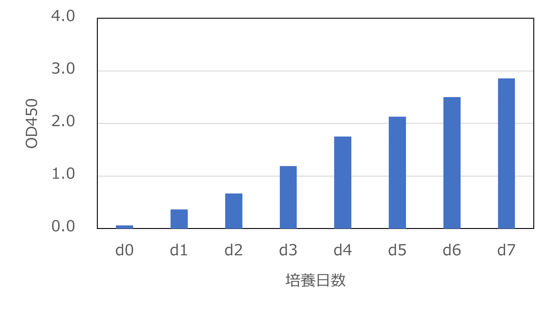
Figure 1. Time course of CD90/CD63-positive exosomes in
MSC culture supernatant UE6E7T-3 cells, which are immortalized MSCs, were seeded in a 12-well dish at 1.0×10 4 cells/cm 2 in 1 mL of POWEREDBY10 medium . The medium collected from each well each day was centrifuged at 2,000xg for 5 minutes to remove debris, frozen, and then measured using this kit without dilution.
- J. Skog, T. Wurdinger, S. van Rijn, DH Meijer, L. Gainche, M. Sena-Esteves, WT Jr. Curry, BS Carter, AM Krichevsky and XO Breakefield: Nat Cell Biol ., 10 , 1470 (2008 ).
- T. Pisitkun, RF Shen and MA Knepper: Proc Natl Acad Sci USA ., 101 , 13368 (2004).
- S. Runz, S. Keller, C. Rupp, A. Stoeck, Y. Issa, D. Koensgen, A. Mustea, J. Sehouli, G. Kristiansen and P. Altevogt: Gynecol Oncol ., 107 , 563 (2007) .
- S. Keller, AK Konig, F. Marme, S. Runz, S. Wolterink, D. Koensgen, A. Mustea, J. Sehouli and P. Altevogt: Cancer Lett ., 278 , 73 (2009).
- C. Lasser, V. Seyed AlikhS. Gabrielsson, J. Lotvall and H. Valadi: J Transl Med ., 9 , 9 (2011).
- Y. Naito, Y. Yoshioka, Y. Yamamoto and T. Ochiya: Cell Mol Life Sci ., 74 , 697 (2017).
- A. Zoraida and M. Yanez-Mo: Front Immunol ., 5 , 442 (2014).
- V. Filipe, A. Hawe and W. Jiskoot: Pharm Res ., 27 , 796 (2010).
- S. Rani, AE Ryan, MD Griffin and T. Ritter: Mol Ther ., 23 , 812 (2015).
- V. Filipe, A. Hawe and W. Jiskoot: Pharm Res ., 27 , 796 (2010).
- M. Théry et al .: J Ext Vesicles , 7 , 1535750 (2018).
Mouse anti-human CD63 antibody
Example data

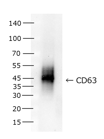
Figure 2. WB analysis of CD63
Sample: Immortalized human mesenchymal stem cell line UE6E7T-3 cell extract (5µg/lane) Western
blotting was performed using this antibody (mouse anti-human CD63 monoclonal antibody, clone No. A1-9A2) as the primary antibody and HRP-labeled anti-mouse IgG antibody as the secondary antibody.
| Product Specifications | |
| Clonality | Monoclonal (Clone No.: A1-9A2) |


