Staining fat globules with BODIPY®
Adipocyte fluorescent staining kit
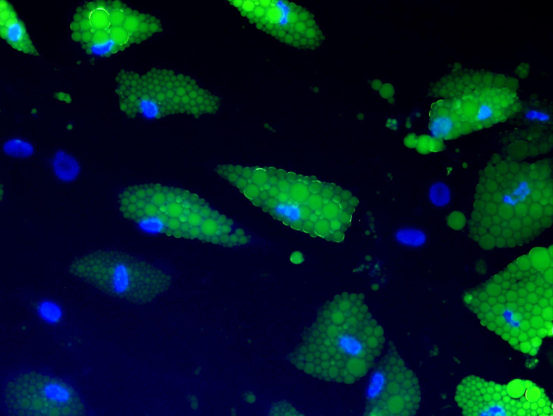
The Adipocyte Fluorescent Staining Kit is an adipocyte staining kit that replaces the conventional Oil Red O staining method. This kit stains intracellular fat globules with BODIPY®* and nuclei with H33258.
Intended use
This kit is an adipocyte staining kit that replaces the conventional Oil Red O staining method . This kit stains intracellular fat globules with BODIPY® * and nuclei with H33258.
This kit is a fat staining kit developed as an optional kit for the separately sold white adipocyte culture kit , brown adipocyte culture kit , and mesenteric adipocyte culture kit. It is possible to quantify the amount and shape of fat per cell using IN Cell Analyzer 1000 (GE Healthcare Bioscience Co., Ltd.).
*BODPY® is a registered trademark of Invitrogen Corporation.

human adipocyte
Composition
- Cleaning liquid (tablet) x 5 for 100 mL
- Fat globule staining solution 50 mL x 1 bottle
- Nuclear staining solution 50 mL x 1 bottle
- Mounting medium 50 mL x 1
* Can be used for 10 96-well plates.
* This kit does not include a fixative (10% neutral buffered formalin). Please purchase separately or make your own according to the attached manual.
Dyeing example

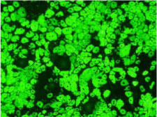
Figure 1: Observation of fat globules
Photographed with excitation light (493 nm) and fluorescence (503 nm)

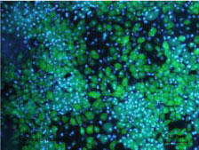
Figure 2: Observation of nuclei
Photographed with excitation light (352 nm) and fluorescence (461 nm)
| Catalog Number | Product Name | Size |
| PMC-AK19F | Adipocyte Fluorescent Staining Kit | 1 Set |
