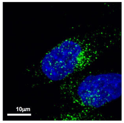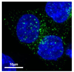
Fluorescent immunostaining of exosomes produced by cancer cells using CD63 antibody (clone: 8A12)
User Report
Nobuyoshi Kosaka
National Cancer Center Research Institute of Molecular and Cellular Therapy

Products
- Anti CD63 for Exosome Isolation, Human (Mouse) Unlabeled, 8A12(Cat.No. SHI-EXO-M02)
Experimental findings
Exosomes are small extracellularly secreted lipid bilayer-delimited vesicles of approximately 100 nm in diameter. The ISEV (International Society for Extracellular Vesicles) recommends the use of the term extracellular vesicles (EVs) to refer to the heterogenous class of secretory vesicles that includes exosomes derived from multivesicular endosomes. Recent exosomes studies have begun to clarify the role of exosomes in some diseases. However, to understand the production mechanism of exosomes it is necessary to have: 1) physiological understanding of the role of exosomes; 2) understanding of the diseases that arise from exosomes. Elucidating the mechanism of exosome secretion by cancer cells may lead to new treatments for cancer.
To track the kinetics of exosome production we performed immunostaining experiments with an anti-human CD63 antibody (clone 8A12; available from Cosmo Bio USA). Using immunofluorescence microscopy, we observed a number of CD63 positive granule-like structures in prostate cancer cell line PC-3M (Figure 1) and in colon cancer cell line HCT116 (Figure 2). In the future we will co-stain cancer cells with anti-human CD63 antibody (clone 8A12) and antibodies for other endosomal markers to explore the production of exosomes by cancer cells. The high specificity of anti-human CD63 antibody (clone 8A12) allowed it to be used successfully to target exosomes in vivo to suppress cancer metastasis.
Figures
 Figure 1. Localization of CD63 in prostate cancer cell line PC-3M
Anti-human CD63 antibody (clone 8A12) was detected by Alexa Fluor™ 488-labeled anti-mouse secondary antibody.
 Figure 2. Localization of CD63 in colon cancer cell line HCT116
Anti-human CD63 antibody (clone 8A12) was detected by Alexa Fluor™ 488-labeled anti-mouse secondary antibody
