Exosome Tetraspanin Monoclonal Antibodies
Exosome monoclonal antibodies specifically recognize CD9, CD63, and CD81 known as exosome markers. They are also antibodies that can isolate exosomes from serum and culture supernatant by immunoprecipitation. *Partially patent granted
Background
Exosomes are vesicles with diameters of 50 nm ~ 150 nm formed by lipid bilayer membranes secreted from cells. They are observed in body fluids such as saliva, blood, urine, amniotic fluid, and malignant ascites, and are also secreted from cultured cells in vivo. In recent years, it has been reported that exosomes contain various proteins and RNA, and may play an important role in transmitting information between cells.
Features
- Recognizes exosome membrane proteins CD9, CD63, CD81 with high specificity.
- Compatible samples (validated with human samples)
CD9: Serum, plasma, culture supernatant, urine
CD63: Serum, plasma, culture supernatant, urine
CD81: Serum, plasma, culture supernatant - * CD9 is bovine milk exosomes, CD81 is bovine milk exosomes, and FBS has also been detected
- Useful for exosome surface antigen protein, endogenous RNA (miRNA), and protein analysis.
Exosome monoclonal antibody Anti CD9
| Catalog Number | Product Name | Size |
| CAC-SHI-EXO-M01-50 | Anti CD9 Antigen (MRP-1/Tspan-29) mAb (Clone 12A12) | 100 ul |
| CAC-SHI-EXO-M01-B | Anti CD9 Antigen (MRP-1/Tspan-29) mAb (Clone 12A12, Biotin Labeled) | 100 ul |
| CAC-SHI-EXO-M01-TF2 | Anti CD9 Antigen (MRP-1/Tspan-29) mAb (Clone 12A12, TF2WS Labeled) | 100 ul |
| CAC-SHI-EXO-M01-TF5 | Anti CD9 Antigen (MRP-1/Tspan-29) mAb (Clone 12A12, TF5 Labeled) | 100 ul |
Exosome Monoclonal Antibody Anti CD63 xxxx
| Catalog Number | Product Name | Size |
| CAC-SHI-EXO-M02-50 | Anti CD63 Antigen (LAMP-3/Tspan-30) mAb (Clone 8A12) | 100 ul |
| CAC-SHI-EXO-M02-B | Anti CD63 Antigen (LAMP-3/Tspan-30) mAb (Clone 8A12, Biotin Labeled) | 100 ul |
| CAC-SHI-EXO-M02-TF2 | Anti CD63 Antigen (LAMP-3/Tspan-30) (Clone 8A12, TF2SW Labeled) | 100 ul |
| CAC-SHI-EXO-M02-TF5 | Anti CD63 Antigen (LAMP-3/Tspan-30) (Clone 8A12, TF5 Labeled) | 100 ul |
zzzzExosome monoclonal antibody Anti-CD81
| Catalog Number | Product Name | Size |
| CAC-SHI-EXO-M03-50 | Anti CD81 Antigen (TAPA-1/Tspan-28) mAb (Clone 12C4) | 100 ul |
| CAC-SHI-EXO-M03-B | Anti CD81 Antigen (TAPA-1/Tspan-28) mAb (Clone 12C4, Biotin Labeled) | 100 ul |
| CAC-SHI-EXO-M03-TF2 | Anti CD81 Antigen (TAPA-1/Tspan-28) mAb (Clone 12C4, TF2SW Labeled) | 100 ul |
| CAC-SHI-EXO-M03-TF5 | Anti CD81 Antigen (TAPA-1/Tspan-28) mAb (Clone 12C4, TF5 Labeled) | 100 ul |
IP-Western Blot
- IP-WB of serum exosome by CD9 antibody 12A12
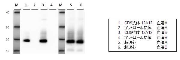
- IP-WB of serum exosome by CD63 antibody 8A12

- IP-WB of serum exosome by CD81 antibody 12C4

Immunofluorescent cell staining (ICC/IF)
| A. | B. |
 |
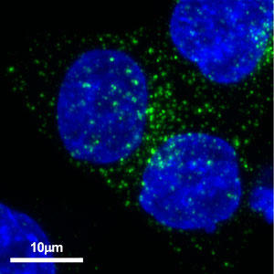 |
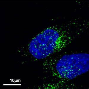
Localization of CD63 in prostate cancer cell PC-3M and colon cancer cell HCT116
A: Prostate cancer cell PC-3M
B: Colon cancer cell HCT116
secondary antibody with Alexa Fluor® 488-conjugated anti-mouse secondary antibody.
Immunoelectron microscopy
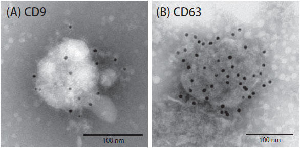
Detection of human CD9 and CD63 proteins on the surface of exosomes by immunoelectron microscopy
(A) Using an anti-human CD9 antibody (clone 12A12), CD9 present on the surface of exosomes derived from human breast cancer cells was detected.
(B) Anti-human CD63 antibody (clone 8A12) was used to detect CD63 present on the surface of exosomes derived from human breast cancer cells.
cryo-electron microscopy
| A. | B. |
 |
 |
C.
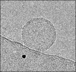
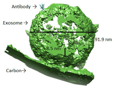
Exosomes and anti-human CD63 antibody (clone 8A12) were mixed and photographed with a cryo-electron microscope, Titan Krios (A).
(B) is a three-dimensional construction of the captured image, and (C) is a rotating movie.
List of Related Application Notes
- Examples of using monoclonal antibodies that recognize exosome markers
- Immunoelectron microscope observation of exosomes using anti-exosome antibodies
- Fluorescent immunostaining of exosomes produced by cancer cells using CD63 antibody (clone: 8A12)
- Detection of exosomal proteins by Western blotting using anti-CD81 antibody
Literature
- ■CD9
- S Tsuda et al., Sci Rep. 2017 Oct 11;7(1):12989. doi: 10.1038/s41598-017-13154-0.
- N Nishida-Aoki et al., Mol Ther. 2017 Jan 4;25(1):181-191. doi: 10.1016/j.ymthe.2016.10.009.
- 2017 Apr 11; 8(15): 24668-24678. doi:10.18632/oncotarget.14969
- Kazutoshi Fujita et al., Sci Rep. 2017; 7: 42961. doi: 10.1038/srep42961
- Yoshioka Y et al., Nat Commun. 2014 Apr 7;5:3591. doi: 10.1038/ncomms4591.
- Saito S et al., Sci Rep. 2018 Mar 5;8(1):3997. doi: 10.1038/s41598-018-22450-2.
- Yagi Y et al., Neurosci Lett. 2017 Jan 1;636:48-57. doi: 10.1016/j.neulet.2016.10.042. Epub 2016 Oct 22.
- Ueda K et al., Sci Rep. 2014 Aug 29;4:6232. doi: 10.1038/srep06232.
- Tokuoka, SM et al. Lipid Profiles of Human Serum Fractions Enhanced with CD9 Antibody-Immobilized Magnetic Beads. Metabolites 12, 230 (2022).
- ■CD63
- N Nishida-Aoki et al., Mol Ther. 2017 Jan 4;25(1):181-191. doi: 10.1016/j.ymthe.2016.10.009.
- Yoshioka Y et al., Nat Commun. 2014 Apr 7;5:3591. doi: 10.1038/ncomms4591.
- Saito S et al., Sci Rep. 2018 Mar 5;8(1):3997. doi: 10.1038/s41598-018-22450-2.
- ■CD81
- M Somiya et al., J Extracell Vesicles. 2018 Feb 21;7(1):1440132. doi:10.1080/20013078.2018.1440132. eCollection 2018.
- Takahashi A et al., Nat Commun. 2017 May 16;8:15287. doi: 10.1038/ncomms15287.

